About Eye Anatomy
Understanding the anatomy of the eye is essential to comprehending cataracts, how and why they impair vision, and cataract surgery in general. Although the human eye is tiny compared with other organs in the body, it contains a number of distinct anatomical components that all contribute to normal sight in some manner.
We All Deserve To See The World with Clarity.
The eye, like a camera, functions on the principle of a shutter. A camera’s shutter can shut or open depending on the amount of light required to expose the film in the back of the camera. The eye and its pupils work in much the same way as a camera’s shutter. Our pupils get bigger when it is very dark so that we can absorb more light. The pupils get smaller in bright light to protect us from too much light exposure.
The eye is a complex organ that is essential for vision. Though we take our eyesight for granted, it is an amazing feat of anatomy and physiology. The eye allows us to see the world around us by focusing light onto the retina, which converts the light into electrical impulses that are sent to the brain. The eye is constantly adjusting the amount of light it takes in, depending on the level of illumination. In low light, the pupil dilates to allow more light in; in bright light, the pupil constricts to protect against too much light exposure.
Book Free Consultation
Book Appointment or Video Consultation online with top eye doctors
Eye Anatomy
The eye’s basic and fundamental anatomy comprises of:
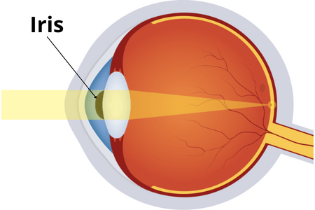
Iris
The iris is the part of the eye that determines its hue, such as blue, brown, green, or hazel. It serves as a diaphragm for a camera and expands (dilates) and contracts to allow more or less light into the eye.
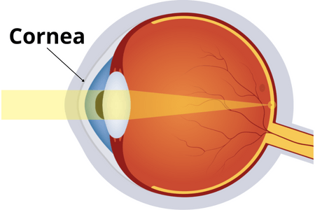
Cornea
The cornea is the eye’s transparent window. It’s located in front of the colored part of the eye, just behind the iris, and it has no blood vessels. It typically survives intact because to its lack of blood vessels. When a person gets a “scratch” to their eye or sustains a corneal abrasion, it is very painful because the cornea has a high concentration of nerve endings.
A corneal transplant is a surgical procedure that may be used to treat individuals with permanent corneal scarring. When people wear contact lenses, they rest on top of the cornea. The cornea is the part of the eye that is laser-scribed during LASIK or PRK procedures to help patients see without glasses (Refractive Surgery).
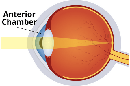
Anterior Chamber
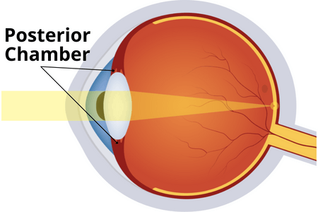
Posterior Chamber
The posterior chamber contains the ciliary body muscle and the lens complex, which is located behind the iris. This chamber produces water, which flows to the front of the eye for drainage into the anterior chamber angle.
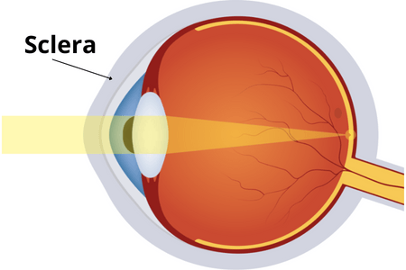
Sclera
The sclera, also known as the white of the eye, is a tough, white fibrous membrane that covers most of the eyeball. It protects and gives shape to the eye.
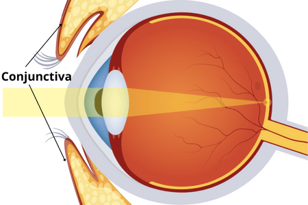
Conjunctiva
The conjunctiva is a transparent membrane that lies on top of the sclera. It covers the eyeball up to the lids and then folds beneath them, covering the underside of both upper and lower lids. A contact lens can never be misplaced behind the eye since the conjunctiva acts as an natural barrier. When we have an infection or an allergy reaction of the eyes, the conjunctiva becomes inflamed and crimson, giving us “pink eyes.” Occasionally, individuals get a red spot beneath the conjunctiva that is a little of blood, which can appear on its own or as a result of minor injury and generally goes away in a few days.
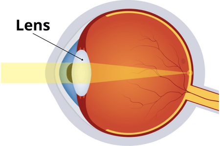
Lens
The natural lens is what you’re seeing through. The clarity of this lens is generally excellent throughout most of our lives, and it changes form as we try to focus on close objects or far-away ones. Our lenses’ ability to alter shape may be temporarily (or permanently) hampered by some medications or illnesses.
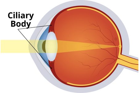
Ciliary Body and Ciliary Muscle
This is the muscle that allows the iris to shift back and forth, shaping the pupil. It also generates eye fluid known as “aqueous.” Diode laser treatment for glaucoma targets the ciliary body to decrease aqueous production, reduce fluid accumulation, and lower pressure in the eye.
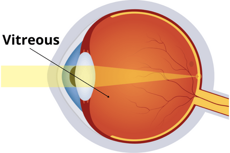
Vitreous
The vitreous is a clear gel that fills the majority of the eyeball. It’s solid when we’re born, but it later detaches from the eye’s interior and becomes loose. “Floaters” are formed as a result of this looser vitreous condition. These are simply pieces of looser vitreous floating in more gel-like vitreous. Sometimes, the gel can form clumps that look like cobwebs.
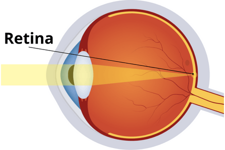
Retina
The retina is the “film” of the eye, much like a camera lens. It’s just a thin layer of nerve tissue covered by a transparent protective covering. If this layer detaches from the circulation and becomes detached, it is an eye emergency that requires surgical treatment to avoid permanent vision loss.
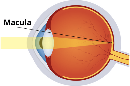
Macula
Macula is a yellowish spot near the center of the retina, the sensitive tissue at the back of the eye. The macula provides clear central vision. It is responsible for what we see directly in front of us. We use our central vision for seeing objects clearly and for doing fine, detailed work, such as reading and sewing.
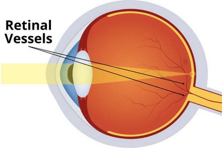
Retinal Vessels
Retinal Vessels are the blood vessels located in the retina of the eye that are responsible for supplying oxygen and nutrients to the retina. The retina is a thin layer of tissue at the back of the eye that is responsible for sensing light and sending signals to the brain. The retinal vessels are very important for maintaining healthy vision.
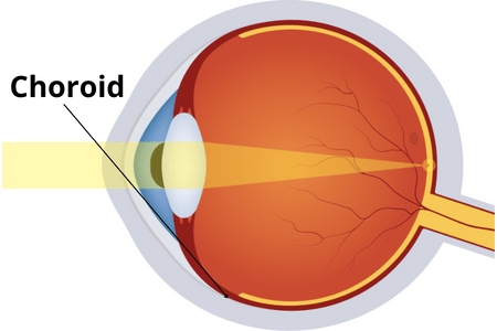
Choroid
The choroid is the layer of the eye that provides most of the blood supply to the retina. It is a thin, dark membrane that lies between the retina and the white outer coat of the eye (the sclera). The choroid helps to keep the eye healthy by providing nutrients and oxygen to the retina.

Optic Nerve
The optic nerve is a fiberoptic cable that connects all of the retinal receptor cells and sends them to the brain for analysis. Our “super computer,” the brain, translates these picture messages for us. Over time, the optic nerve deteriorates in glaucoma, losing this essential link to the brain. The result is loss of vision.